Skip to main content
We help people live a pain-free, active and happy life.
Home
About
Doctors Unscripted Podcast
LIVE A LIFE PAIN-FREE
Select the location nearest you.
Arizona
Ohio & Indiana
As Featured In National Media
Our innovative treatments, world-class physicians, and disruptive approach to pain relief has garnered national attention.
Advanced Procedures
We pair exceptional care by world-class physicians with innovative treatments driven by proven, cutting-edge technology.
Peripheral Nerve Stimulation (PNS)
MILD
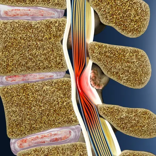
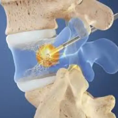
Intracept
Spinal Cord Stimulation (SCS)
View All Projects
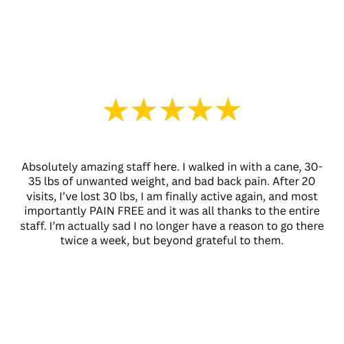
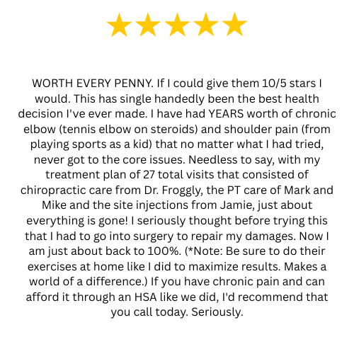
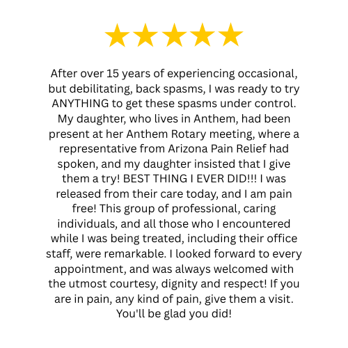
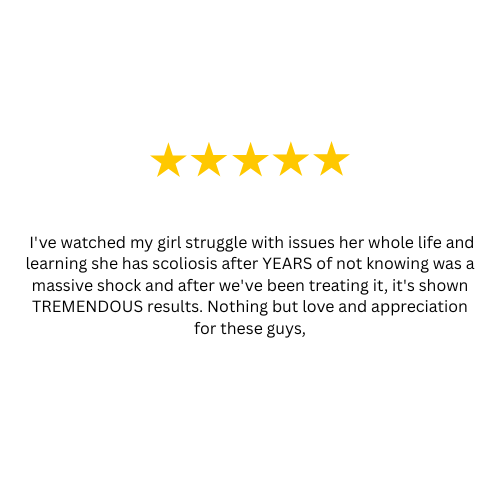
Patient Testimonials